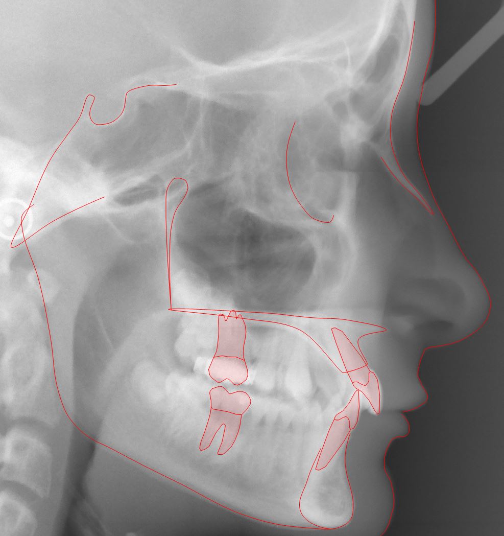
Tracing
Tracing is the placement of graphic symbols on a digital tracing image, most likely a lateral x-ray image. Anatomic structures to be traced are hard and soft tissue landmarks (markers), teeth, hard tissue segments, a soft tissue profile line, and other structures.
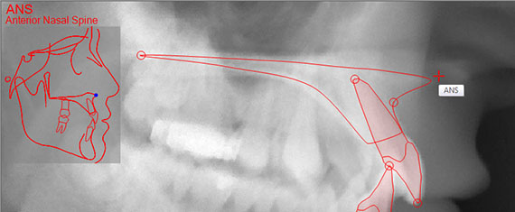
Landmarks (markers)
Facad enables a quick and easy method to interactively place markers directly on the x-ray image with just a simple mouse-click.
You will be guided throughout the process with the help of a placement guide that shows the correct position for a landmark, allowing you to place the markers necessary for the chosen analysis.
The mouse cursor is also positioned automatically, to the vicinity of the correct position.
Any marker can afterwards be adjusted in order to be accurately placed.
The zoom function enables you to place the markers with great accuracy.
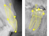
Teeth
Predefined graphic symbols for incisors are automatically drawn when the incisors’ markers are placed.
Symbols for canines, premolars, and molars are interactively placed directly on the x-ray image with two clicks of the mouse.
A placed tooth can later be adjusted regarding its position, size, and/or slope.
Hard tissue
Hard tissue segments such as the maxilla and the mandible, and other anatomical structures are placed/drawn on the x-ray image using ready-made templates.
The placed hard tissue segments can later be adjusted.
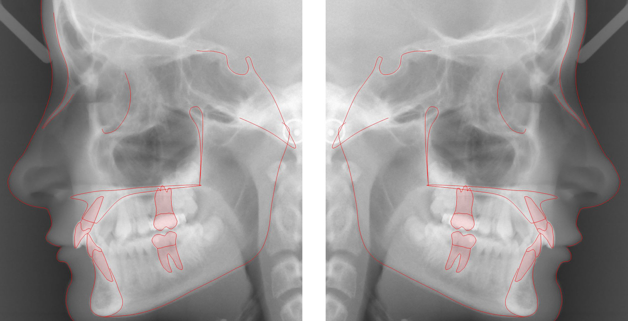
Soft tissue profile line
The soft tissue profile line is placed/drawn on the x-ray image using a predefined template and with the aid of automatic structure recognition.
The profile line can afterwards be adjusted.