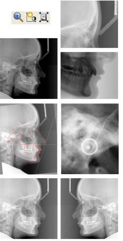
Facad www.facad.com

Images
Tracing, cephalometric analysis, and treatment planning is performed in Facad with the use of digital images. A digital x-ray image can either be a scanned x-ray film or a digital image file in JPEG, TIFF, BMP, or DICOM format.

Zoom
An image can be enlarged (zoomed) so that structures in the image can be scrutinized and markers (landmarks) can be placed very accurately.
This is easily accomplished with the scroll-wheel of the mouse or with a key on the keyboard.


Brightness and contrast
The brightness and contrast of an image can interactively be adjusted.

It is possible to optimise the brightness and contrast for a certain area of the image.

The image can also be inverted.


Mirror and rotate
An image can be rotated and mirrored in order to have it correctly displayed.

It is also possible to freely rotate the image.


Plugin programs
Facad is adapted to be able to receive patient data and digital images from other software such as programs for digital x-ray imaging, patient management systems, or PACS.

The following list shows some examples of systems and software that Facad already is adapted to:

  • Dimaxis (Planmeca)
  • Romexis (Planmeca)
  • Sidexis (Sirona)
  • CliniView (Instrumentarium Imaging)
  • Digora (Soredex)
  • CDR Dicom (Schick)
  • Vixwin (Gendex)
  • DBSwin (Dürr)
  • Kodak Dental Imaging Software
  • Effica Viewer (Tieto)
  • IDS5 PACS (Sectra)
  • IDS7 PACS (Sectra)
  • iSite Enterprise PACS (Philips)
  • PACS systems with Dicom Send abilities (Facad = Image Storage SCP)
  • VisiQuick (Thomas Monitor Systems)
  • OrthoPLAN (FIS Automatisering)
  • Titanium (Flexsoft)
  • DentalEye
  • Edward/Paul/Arion (ProCuris)
  • T4 (Kodak)
  • Opus Dental
  • Orthodontis
  • TK2 (DSI Data)
  • + any software that can export images to image files in Dicom format.